Photographing River Mussels for iNaturalist
Walk through any river or along any lakeshore, and you might be fortunate to spot a River Mussel (Order: Unionida). If you'd like to know what species of mussel you're looking at you might upload it to iNaturalist to help find some answers. Mussels are incredibly diverse so it's essential to photograph the correct angles. However, it may not always be intuitive to know which features you should photograph.
I've put together a brief primer on mussel terminology and highlighted important features and angles to photograph so that we can make the most of your mussel observations!
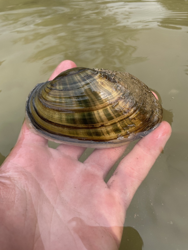
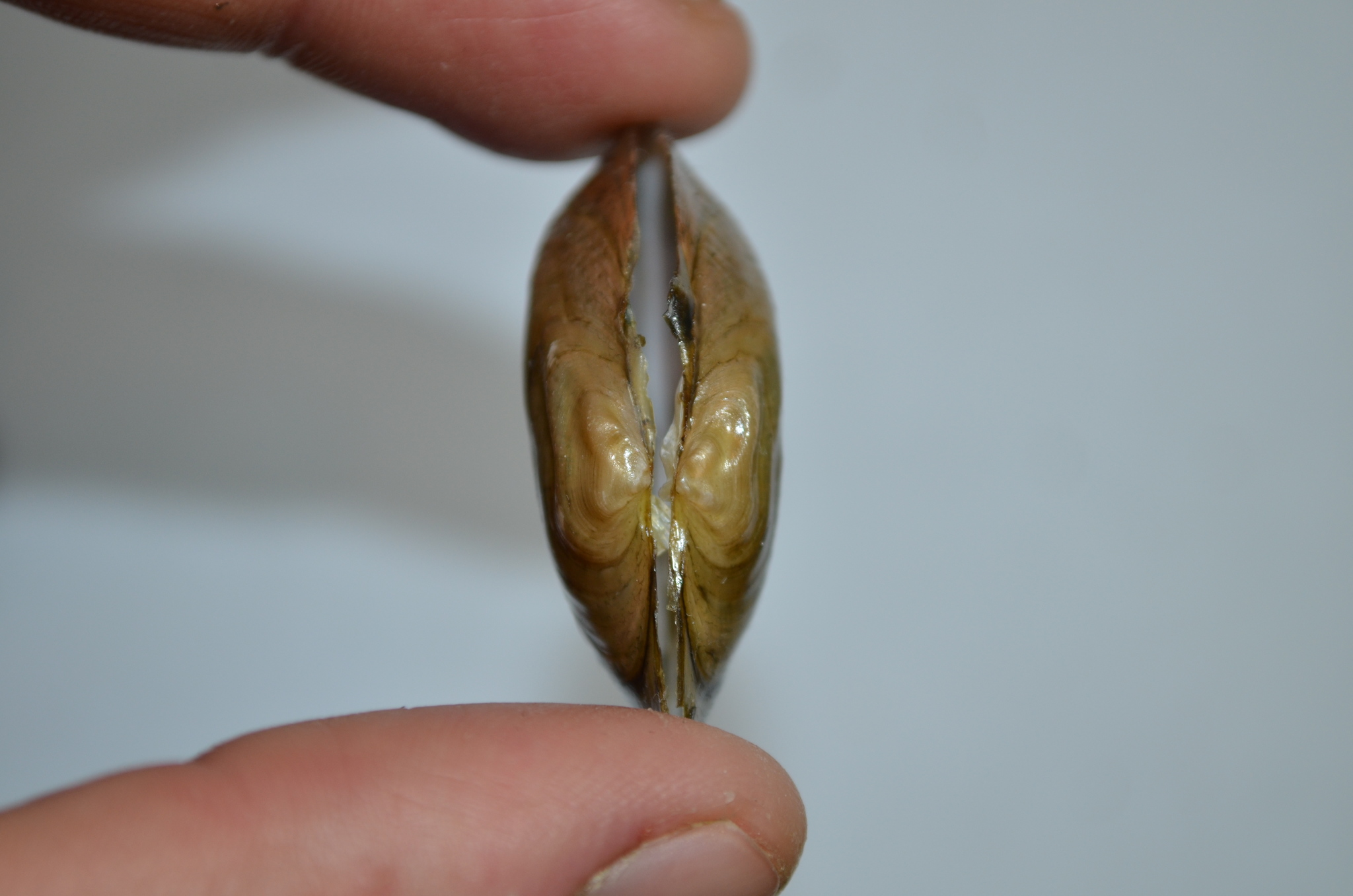
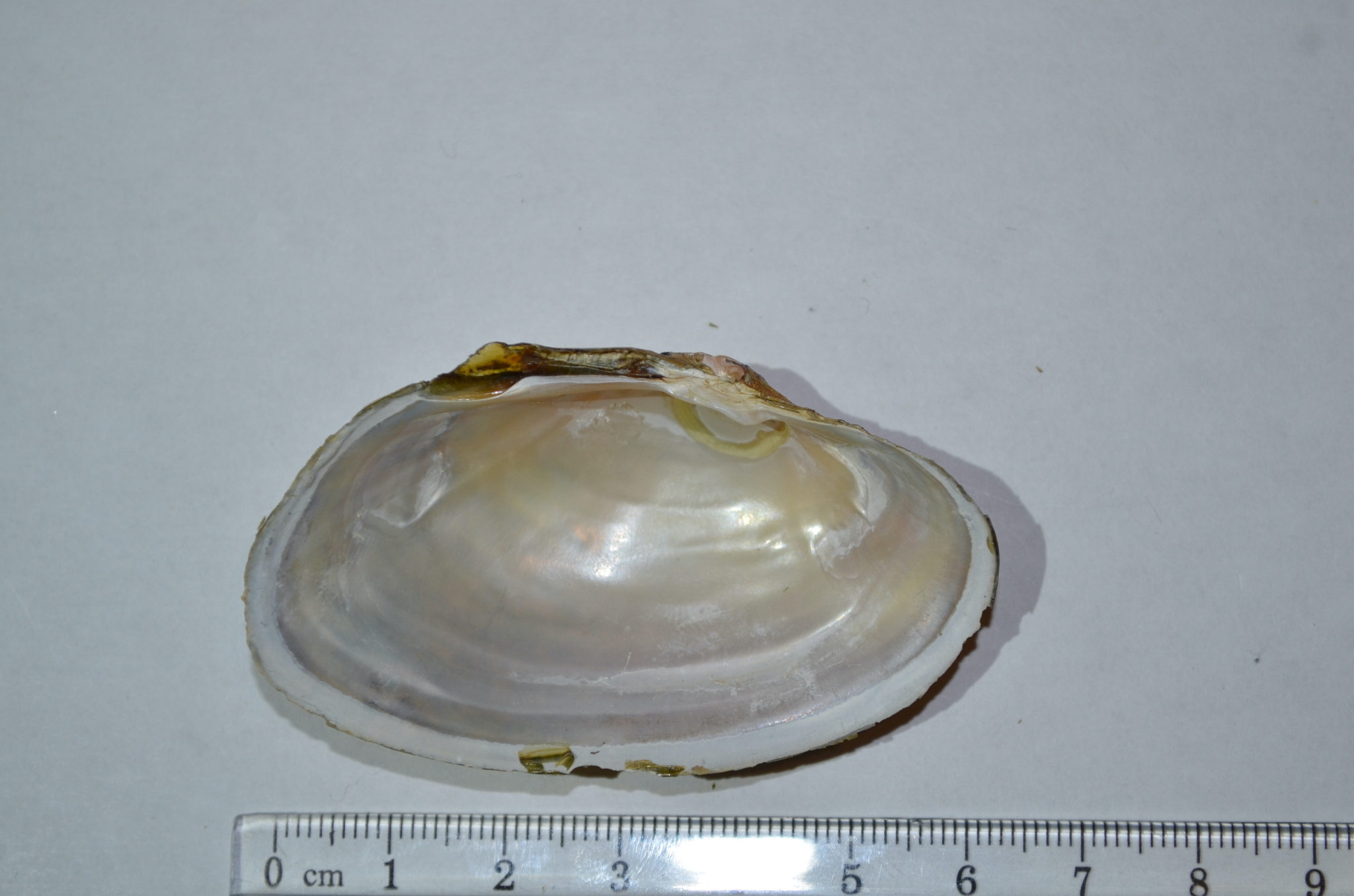
Here is a lateral photo of a typical River Mussel. Its dorsal is at the top, ventral at the bottom, anterior on the left, and posterior on the right. That makes the side facing us the left valve.
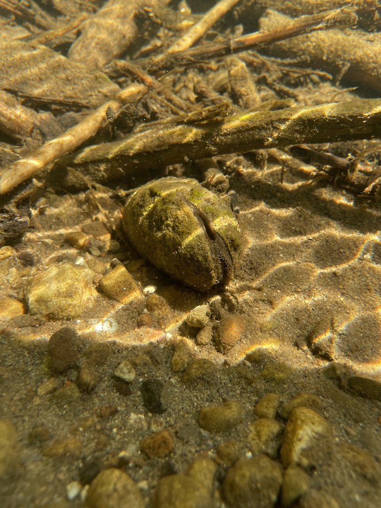
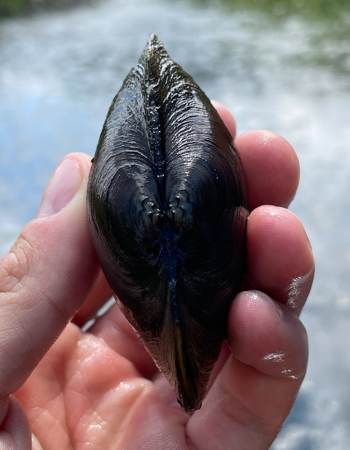
Here is a River Mussel in situ. Its posterior is exposed with its exhalant siphon above and inhalent siphon below. The anterior of the mussel typically stays buried and is secured by a muscular foot. *An in situ photo is not required for identification.
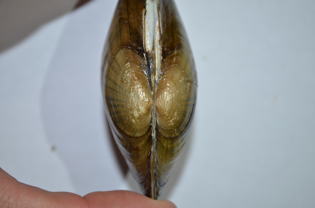
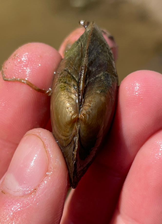
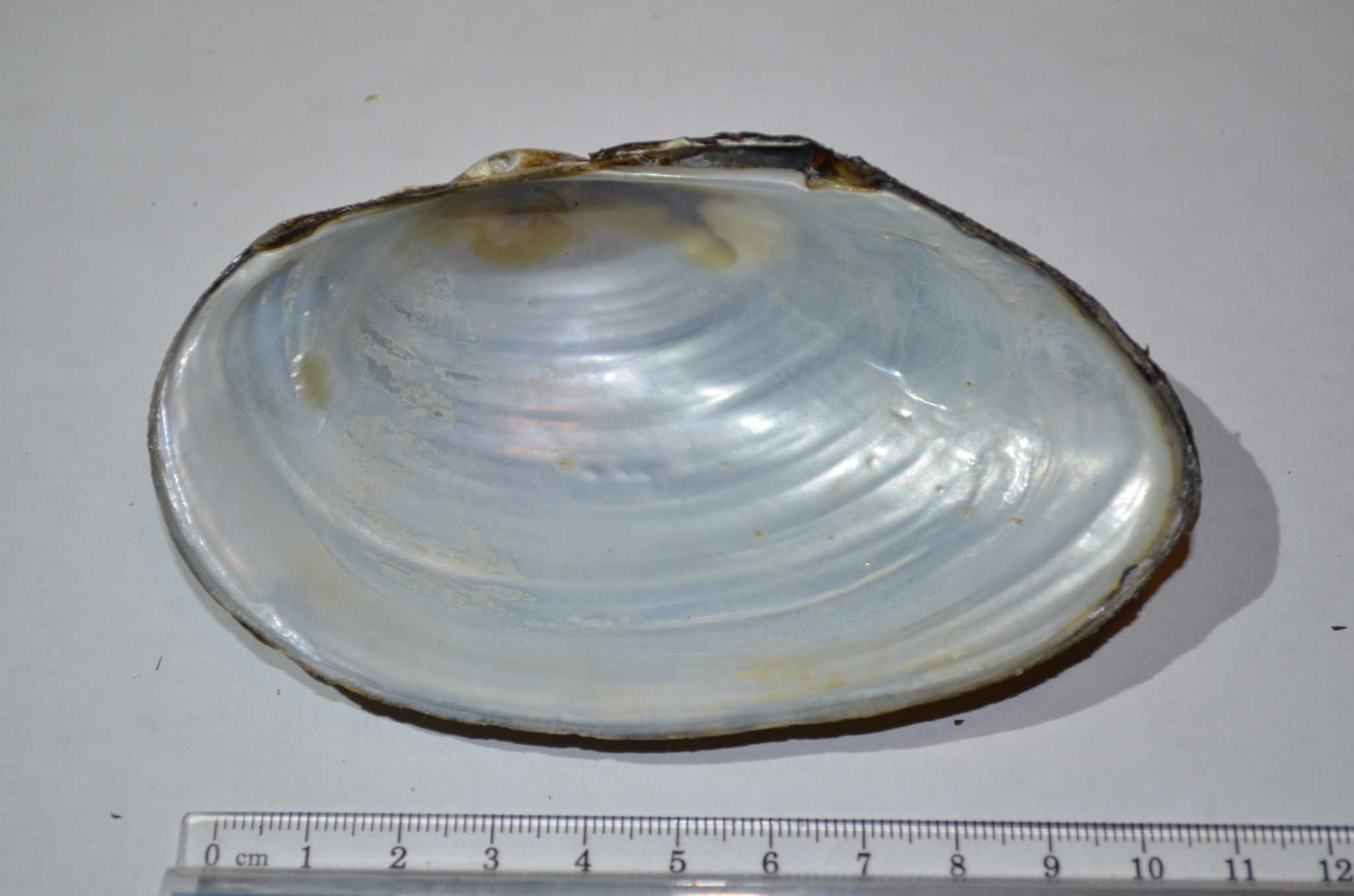
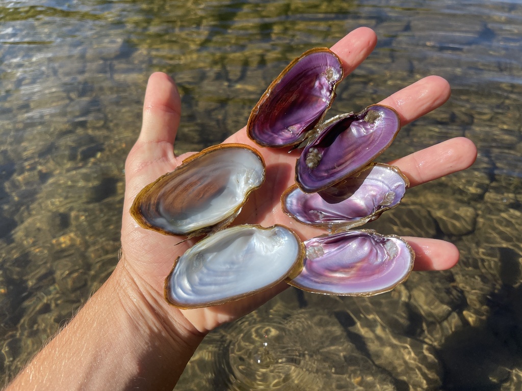
From the exterior of the mussel, there are two important angles to photograph: a Lateral Photo (see first photo), and a Dorsal Photo of the Beak Sculpture. The lateral photo will show the profile of the mussel, as well as the exterior colour and patterning. The exterior surface is called the Periostracum. The Beak is the oldest part of the shell where the two valves come to a point. Beak sculpture refers to the raised contours formed in the first year of a mussel's growth, it can be diagnostic for some species. Below I've included four examples to illustrate the diversity of beak sculptures between species. Before taking a photo of the beak sculpture it can be helpful to clean the area off of any sediment and river deposition.
Fine, wavy lines
Sharp, single-loop ridges
Heavy, double-loop bars
Interrupted, nodulous double-loop ridges
Above the beak is the Ligament. This is the hinge on the mussel and it is always posterior to the beak. The ligament forms the Hinge Line of the mussel.
The interior often holds the most important characters for identification, but unfortunately, they're only available when a mussel is dead. Inside are "teeth", these are not for chewing but to interlock and help hold both valves together securely. A typical mussel will have "complete" teeth, meaning that both pseudocardinal teeth and lateral teeth are present. The Pseudocardinal Teeth sit beneath the beak towards the anterior. The Lateral Teeth follow the hinge line towards the posterior. Below I've included some examples of differences in teeth between species.
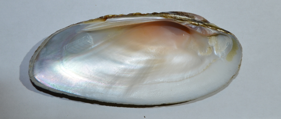
Complete dentition: Lateral teeth and pseudocardinal teeth present
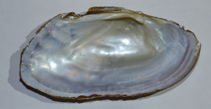
Incomplete dentition: Lateral teeth vestigial (reduced to flattened thickening), pseudocardinal teeth present
Incomplete dentition: Lateral and pseudocardinal teeth vestigial. Lateral tooth reduced to thickening along hinge line, pseudocardinal reduced to protruding knob
Hinge teeth absent: No thickening or other indication of teeth
Hopefully, we can all speak the same language now and you know a little more about mussels. I look forward to seeing your next observations!
Happy musseling,
Sam Turner


